What is the difference between the radiological and clinical picture of a patient

The Schroth method is a traditional method used in the non-operative treatment of scoliosis and other spinal deformities. The benefits of applying the Schroth method are manifold. Among others, improvement of the clinical status can be achieved in children who are in the phase of growth and development.
What is a clinical image and what is a radiological image and what is the difference are just some of the questions you often ask us.
The clinical picture or clinical status represents all the changes in the child’s body that occurred as a result of the deformity. The clinical picture is determined on the basis of a visual examination. The most frequent changes that can be observed on the child’s body are a displaced and rotated pelvis, one hip that is at a higher level than the other, then the existence of the so-called rib gibbus, i.e. the ribs on one side of the chest are more pronounced and rotated backwards, also quite often uneven shoulder level is also present.
When bending the trunk forward (Adam’s test), a more pronounced side of the trunk in the thoracic or lumbar part can be observed, which is created as a result of the rotation of the spinal column.
Corrective exercises improve the clinical picture, which primarily means bringing the body back into balance. Observing the child, we return the pelvis displaced from the belt to the central position, and the chest and shoulders are brought to a more symmetrical position.
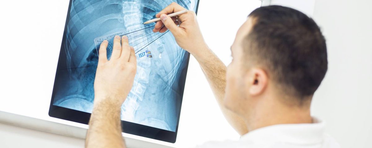
The radiological image represents the characteristics of the spinal column that can only be seen on an X-ray. Using a radiological image, it is possible to determine the size of the scoliotic curve according to Cobb, rotation of the vertebrae, balance, degree of bone maturity and other parameters that cannot be determined by visual examination. When we talk about the radiological result, we mean a reduction in the size of the curve, a reduction in the rotation of the vertebrae, which can only be determined using an X-ray.
